Blog

  • Laser Photobiomodulation in Dentistry: Revolutionizing Healing and Pain Management

    Laser Photobiomodulation in Dentistry: Revolutionizing Healing and Pain Management

    Introduction

    Modern dentistry has evolved far beyond simply treating cavities and replacing missing teeth. Today, dental care focuses not only on achieving successful clinical outcomes but also on improving comfort, reducing recovery time, and enhancing the overall treatment experience. As technology continues to advance, dental professionals are increasingly adopting innovative solutions that support healing while minimizing discomfort.

    One such advancement is Laser Photobiomodulation (PBM), also known as Low-Level Laser Therapy (LLLT). This non-invasive treatment uses specific wavelengths of low-level laser light to stimulate the body’s natural healing processes. Unlike traditional surgical lasers that cut or remove tissue, PBM works at a cellular level to encourage repair, reduce inflammation, and support recovery.

    Over the past few years, photobiomodulation has gained significant attention across various dental specialties, including implant dentistry, oral surgery, orthodontics, periodontics, and pain management. Its ability to enhance healing while providing a comfortable treatment experience makes it a valuable addition to modern dental care.

    As more individuals seek treatments that are effective, minimally invasive, and recovery-friendly, Laser Photobiomodulation is becoming an increasingly important part of contemporary dentistry.

    What is Laser Photobiomodulation?

    Laser Photobiomodulation is a therapeutic procedure that uses low-intensity laser light to stimulate biological activity within tissues. The treatment is designed to support the body’s natural healing mechanisms and promote healthier tissue function.

    Unlike high-powered surgical lasers that are used to cut, remove, or reshape tissues, PBM lasers deliver gentle light energy without producing significant heat. This means the procedure is comfortable, non-invasive, and does not damage surrounding tissues.

    When the light reaches the targeted area, it interacts with cells and encourages them to perform their repair functions more efficiently. This increased cellular activity can help improve circulation, support tissue regeneration, and reduce inflammation associated with various dental conditions and procedures.

    Because PBM works by enhancing natural biological processes rather than masking symptoms, it is often used as a supportive therapy alongside many dental treatments. The goal is not only to address dental concerns but also to create a smoother and more comfortable recovery experience.

    Today, Laser Photobiomodulation is widely recognized as an advanced technology that complements modern dental care and contributes to better treatment outcomes.

    How Does Photobiomodulation Work?

    Every cell in the human body requires energy to function, repair itself, and maintain healthy tissues. Laser Photobiomodulation works by delivering light energy to targeted tissues, encouraging cells to become more active and efficient.

    When the laser light penetrates the treatment area, it stimulates cellular processes that are essential for healing and regeneration. As cellular activity increases, tissues receive greater support for repair and recovery.

    This process may contribute to:

    • Improved blood circulation
    • Enhanced oxygen delivery to tissues
    • Increased cellular activity
    • Reduced inflammation
    • Faster tissue regeneration
    • Improved collagen production
    • Greater comfort during healing
    • Support for bone and soft tissue recovery

    These biological responses help create an environment that encourages healing and recovery following dental treatments.

    Rather than simply addressing symptoms, PBM works at the source by supporting the body’s natural ability to repair itself. This is one of the reasons why it has become such a valuable tool in modern dentistry.

    Because of these healing properties, Laser Photobiomodulation is now being incorporated into a wide range of dental procedures to improve patient comfort and support successful treatment outcomes.

    Clinical Applications of Photobiomodulation in Dentistry

    1. Dental Implant Healing

    Dental implants have become one of the most predictable and long-lasting solutions for replacing missing teeth. However, the success of an implant depends heavily on proper healing of both the surrounding bone and soft tissues.

    After implant placement, the body begins a natural healing process in which the implant gradually integrates with the jawbone. During this phase, healthy tissue response is essential for long-term stability and success.

    Laser Photobiomodulation can be used as a supportive therapy during implant healing. By encouraging circulation and promoting tissue recovery, PBM may help create favorable conditions around the implant site.

    Many individuals report greater comfort during recovery when PBM is included as part of their treatment plan. It may also help reduce postoperative swelling and support healthier healing of the gums surrounding the implant.

    Potential benefits after implant placement include:

    ✔ Reduced discomfort

    ✔ Improved soft tissue healing

    ✔ Reduced inflammation

    ✔ Enhanced circulation

    ✔ Support for overall recovery

    As implant dentistry continues to evolve, PBM is becoming an increasingly valuable tool for supporting the healing process.

    2. Wisdom Tooth Extraction and Oral Surgery

    Wisdom tooth removal is one of the most commonly performed oral surgical procedures. While the treatment itself is routine, the recovery period may involve swelling, tenderness, stiffness, and temporary discomfort.

    Laser Photobiomodulation can play an important role in supporting recovery following oral surgery. By encouraging tissue repair and helping control inflammation, PBM may contribute to a smoother healing experience.

    Many dental professionals use PBM after wisdom tooth extractions because it provides additional support without requiring invasive procedures or extended recovery measures.

    Individuals often appreciate the ability to return to their daily activities more comfortably while healing progresses naturally.

    Potential advantages include:

    • Reduced swelling
    • Improved comfort
    • Faster tissue recovery
    • Enhanced healing response
    • Better postoperative experience

    As a result, PBM is becoming an increasingly popular addition to oral surgery care.

    3. Management of Temporomandibular Joint Disorders (TMJ)

    The temporomandibular joint, commonly known as the TMJ, connects the lower jaw to the skull and allows movements required for speaking, chewing, and yawning.

    When problems develop within this joint or the surrounding muscles, individuals may experience symptoms that interfere with daily activities.

    Common signs of TMJ disorders include:

    • Jaw pain
    • Facial discomfort
    • Clicking or popping sounds
    • Muscle tightness
    • Difficulty opening the mouth
    • Headaches associated with jaw tension

    Laser Photobiomodulation may help support TMJ management by encouraging circulation and reducing inflammation within affected tissues. The therapy is comfortable, non-invasive, and does not involve medication.

    Many individuals report reduced muscle tension and improved comfort following treatment. Because of these benefits, PBM is increasingly being incorporated into comprehensive TMJ care plans.

    4. Orthodontic Treatment and Braces

    Orthodontic treatment helps improve alignment, bite function, and overall oral health. Whether using traditional braces or clear aligners, tooth movement naturally creates pressure within the supporting tissues.

    As teeth gradually shift into their new positions, temporary soreness and sensitivity may occur.

    Laser Photobiomodulation can help make orthodontic treatment more comfortable by supporting healthy tissue response throughout the treatment process.

    Potential benefits include:

    • Reduced discomfort after adjustments
    • Improved comfort during treatment
    • Support for healthy gum tissues
    • Enhanced overall treatment experience

    For individuals undergoing long-term orthodontic care, PBM may help create a smoother and more comfortable journey toward achieving a healthier smile.

    5. Treatment of Oral Ulcers

    Oral ulcers can significantly affect daily life. Simple activities such as eating, drinking, speaking, and brushing may become uncomfortable when ulcers are present.

    These sores may develop due to stress, irritation, injury, immune responses, or other underlying factors.

    Laser Photobiomodulation offers a gentle and non-invasive approach to supporting ulcer management. The treatment helps encourage tissue repair while reducing discomfort associated with the affected area.

    PBM may be beneficial for:

    • Aphthous ulcers
    • Traumatic ulcers
    • Recurrent mouth sores
    • Oral mucosal irritation

    Many individuals notice improved comfort and faster healing following treatment, making PBM a valuable option for managing oral soft tissue conditions.

    6. Burning Mouth Syndrome

    Burning Mouth Syndrome is a chronic condition that causes a persistent burning sensation within the mouth, even when no obvious abnormalities are visible.

    Individuals may also experience:

    • Tingling sensations
    • Dryness
    • Altered taste perception
    • Ongoing oral discomfort

    Because the condition can affect eating habits, daily comfort, and overall quality of life, finding effective supportive treatments is important.

    Laser Photobiomodulation is increasingly being explored as a non-invasive option for improving comfort in individuals experiencing Burning Mouth Syndrome. By supporting healthy tissue function and reducing irritation, PBM may help alleviate symptoms and improve overall wellbeing.

    7. Gum Disease and Periodontal Therapy

    Healthy gums provide the foundation for strong teeth and long-term oral health. When gum disease develops, inflammation can gradually affect the tissues that support the teeth.

    Treatment often focuses on controlling infection and restoring healthier gum conditions. Following periodontal procedures, tissue healing becomes an important part of successful treatment.

    Laser Photobiomodulation is frequently used alongside periodontal therapy to support recovery and encourage healthier tissue repair.

    Benefits may include:

    • Reduced inflammation
    • Improved healing response
    • Enhanced tissue regeneration
    • Better comfort after treatment

    As laser technology becomes more integrated into dental care, PBM is helping improve the recovery experience for individuals undergoing gum treatments.

    8. Root Canal Treatment

    Root canal treatment is performed to preserve teeth affected by infection or damage. While the procedure effectively addresses the underlying issue, some individuals may experience temporary tenderness as tissues recover.

    Laser Photobiomodulation can be used after root canal treatment to support healing and improve comfort during recovery.

    By encouraging tissue repair and helping reduce inflammation, PBM may contribute to a more comfortable healing experience.

    Potential benefits include:

    • Reduced tenderness
    • Improved comfort
    • Support for tissue recovery
    • Enhanced healing response

    This additional support can help individuals return to normal activities more comfortably following treatment.

    Advantages of Laser Photobiomodulation

    One of the main reasons for the growing popularity of Laser Photobiomodulation is the wide range of benefits it offers.

    Some of the key advantages include:

    Non-Invasive Treatment

    No cutting, surgical intervention, or tissue removal is required.

    Comfortable Procedure

    The treatment is gentle and generally well tolerated.

    Drug-Free Support

    PBM can help support recovery without relying solely on medications.

    Faster Healing

    The therapy encourages natural tissue repair and regeneration.

    Reduced Inflammation

    Swelling and tissue irritation may be minimized.

    Safe for Various Age Groups

    Suitable for children, adults, and older individuals when clinically appropriate.

    Enhanced Treatment Experience

    Supports a more comfortable recovery process across multiple dental procedures.

    These benefits make PBM a valuable addition to modern dental care.

    Is Photobiomodulation Safe?

    Yes. Laser Photobiomodulation is considered a safe and well-established treatment when performed by trained dental professionals.

    The therapy uses low-level laser energy specifically designed to stimulate healing rather than alter or damage tissues. Unlike surgical lasers, PBM does not cut, burn, or remove tissue.

    The procedure is comfortable, non-invasive, and generally well tolerated by individuals of different age groups. Because it supports natural healing processes, PBM has become a trusted adjunctive therapy across many areas of dentistry.

    As with any dental procedure, treatment should always be performed under professional supervision to ensure appropriate care and optimal outcomes.

    Future of Laser Dentistry

    The future of dentistry is increasingly focused on technologies that improve comfort, reduce treatment-related stress, and support faster recovery. Laser Photobiomodulation aligns perfectly with these goals.

    As research continues to expand, PBM is expected to play a greater role in implant dentistry, orthodontics, oral surgery, periodontics, endodontics, and oral medicine.

    Dental professionals are increasingly recognizing the value of therapies that support natural healing while enhancing treatment experiences. With ongoing technological advancements and growing clinical adoption, Laser Photobiomodulation is likely to become a routine part of modern dental care.

    Its ability to complement existing treatments while improving comfort makes it one of the most promising innovations in contemporary dentistry.

    Why Patients Prefer Laser-Assisted Dentistry

    Today’s individuals are more informed than ever before when choosing dental treatments. They often look for solutions that are effective, minimally invasive, and designed to provide a comfortable experience.

    Laser-assisted therapies are becoming increasingly popular because they offer:

    • Comfortable treatment experiences
    • Reduced recovery time
    • Modern technology-driven care
    • Support for natural healing
    • Less treatment-related discomfort
    • Improved overall experiences

    Photobiomodulation reflects the direction in which modern dentistry is moving—toward treatments that prioritize both clinical success and patient comfort.

    Experience Advanced Laser Dentistry at Digital Bite Multispeciality Dental & Implant Hospital

    At Digital Bite Multispeciality Dental & Implant Hospital, we combine advanced dental technology with personalized care to provide treatment experiences that are comfortable, efficient, and focused on long-term oral health.

    Our laser-assisted therapies are designed to support healing, reduce discomfort, and enhance recovery following various dental procedures. Whether you are undergoing implant treatment, oral surgery, gum therapy, orthodontic care, or post-treatment recovery, our team is committed to providing comprehensive care using modern dental solutions.

    Go Digital & Smile Better

    Digital Bite Multispeciality Dental & Implant Hospital

    📍 ICRISAT Colony Phase-1, Ameenpur Police Station Road, Beeramguda

    📞 8019233440
  • Guided Implantology – The Future of Safe & Precise Dental Implants

    Guided Implantology – The Future of Safe & Precise Dental Implants

    Guided Implantology – The Future of Safe & Precise Dental Implants

    Dental implants have become the gold standard for replacing missing teeth. With advancements in technology, Guided Implantology has taken implant procedures to the next level—making them safer, faster, and more precise.

    What is Guided Implantology?

    Guided Implantology is an advanced dental technique that uses 3D CBCT imaging and computer-guided software to plan and place dental implants with extreme accuracy.

    A custom surgical guide is created to ensure precise implant placement, minimizing risks and improving outcomes.

    How Does It Work?

    • 3D CBCT scan captures detailed images of your jaw
    • Digital software plans the exact implant position
    • A surgical guide is created for precise placement
    • Implant is placed accurately with minimal invasion

    Benefits of Guided Implantology

    • Highly accurate implant placement
    • Reduced surgery time
    • Minimally invasive procedure
    • Faster healing and recovery
    • Lower risk of complications
    • Better long-term success

    Who Can Benefit from This Treatment?

    • Patients with missing teeth
    • Cases requiring high precision
    • Full mouth rehabilitation patients
    • Patients with limited bone structure

    Why It’s Better Than Traditional Implants

    Unlike traditional methods, guided implantology eliminates guesswork. It ensures implants are placed exactly where they should be, leading to predictable and long-lasting results.

    Why Choose Digital Bite for Dental Implants?

    Digital Bite uses advanced CBCT technology, experienced implant specialists, and a prosthetically driven approach to ensure safe and successful implant procedures.

    Conclusion

    Guided Implantology represents the future of dental care—combining technology with expertise to deliver precise, comfortable, and reliable results. If you’re considering dental implants, this modern approach offers the best possible outcomes.

  • What is Full Mouth Rehabilitation? A Complete Guide to Restoring Your Smile

    What is Full Mouth Rehabilitation? A Complete Guide to Restoring Your Smile

    What is Full Mouth Rehabilitation? A Complete Guide to Restoring Your Smile

    A healthy smile is not just about appearance—it plays a vital role in chewing, speaking, and overall confidence. But what happens when multiple teeth are damaged, missing, or worn out? This is where Full Mouth Rehabilitation (FMR) comes in.

    What is Full Mouth Rehabilitation?

    Full Mouth Rehabilitation is a comprehensive dental treatment designed to restore all teeth in the upper and lower jaws. It combines multiple dental procedures such as restorative dentistry, cosmetic treatments, implants, and sometimes surgery to rebuild your smile completely.

    Who Needs Full Mouth Rehabilitation?

    You may need FMR if you have:

    • Multiple missing teeth
    • Severely worn or damaged teeth
    • Chronic jaw pain or bite problems
    • Difficulty in chewing or speaking
    • Advanced gum disease

    Key Benefits of Full Mouth Rehabilitation

    • Restores proper chewing and bite function
    • Enhances facial appearance and smile aesthetics
    • Reduces jaw pain, headaches, and TMJ issues
    • Prevents further dental damage
    • Improves confidence and quality of life

    What Treatments Are Included?

    Depending on your condition, FMR may include:

    • Dental implants
    • Crowns and bridges
    • Veneers
    • Root canal treatments
    • Orthodontic corrections

    Advanced Option: BPS Dentures

    For patients with complete tooth loss, BPS dentures offer:

    • Superior fit and comfort
    • Natural-looking appearance
    • Improved speech and chewing ability
    • Long-lasting durability

    Why Choose Digital Bite for Full Mouth Rehabilitation?

    At Digital Bite, treatments are personalized using advanced digital technology and expert care. The focus is always on comfort, precision, and long-term results.

    Conclusion

    Full Mouth Rehabilitation is not just a treatment—it’s a life-changing transformation. If you are struggling with multiple dental issues, this comprehensive solution can restore both your smile and confidence.

  • Invisalign Aligners Explained: What to Expect at Every Stage

    Invisalign Aligners Explained: What to Expect at Every Stage

    Invisalign Clear Aligners: A Simple and Comfortable Way to Straighten Your Smile

    A straight smile is not just about looking good—it also helps improve your oral health, makes cleaning your teeth easier, and boosts your confidence. Many people avoid getting their teeth straightened because they don’t want to wear noticeable metal braces. Fortunately, modern dentistry offers a better solution: Invisalign Clear Aligners.

    Invisalign is a clear aligner system that gradually moves your teeth into the correct position without the need for metal brackets or wires. Since the aligners are transparent and removable, they fit easily into your daily life, making them a popular choice for both teenagers and adults.

    At Digital Bite Multispeciality Dental & Implant Hospital, Beeramguda, we use advanced digital technology, including 3D Digital Intraoral Scanning and 3D CBCT Imaging, to plan your treatment with precision and deliver predictable, comfortable results.

    What is Invisalign?

    Invisalign is a series of custom-made, clear plastic aligners designed to gently move your teeth into the correct position. Each aligner is specially made for your smile and is replaced every 1–2 weeks as your teeth gradually become straighter.

    Unlike traditional braces, Invisalign aligners are removable, comfortable, and almost invisible.

    Benefits of Invisalign

    • Nearly invisible appearance
    • Comfortable with no metal wires or brackets
    • Easy to remove while eating and brushing
    • No food restrictions
    • Easy to maintain oral hygiene
    • Predictable treatment results
    • Fewer dental visits

    How Does Invisalign Work?

    Your Invisalign treatment follows a simple digital process:

    1. Consultation

    Our dentist examines your teeth, bite, and smile to determine if Invisalign is the right option.

    2. Digital 3D Scan

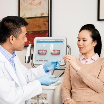
    Using our advanced 3D Digital Intraoral Scanner, we create a highly accurate digital model of your teeth—without messy impression materials.

    3. Personalized Treatment Plan

    A customized digital plan shows how your teeth will move throughout treatment. You can even preview your future smile before you begin.

    4. Custom Aligners

    A series of clear aligners is created specifically for your teeth.

    5. Wear Your Aligners

    Wear your aligners for 20–22 hours every day and change them as advised by your dentist.

    6. Regular Check-ups

    Quick review appointments help ensure your treatment stays on track.

    What Can Invisalign Treat?

    Invisalign can successfully correct:

    • Crooked teeth
    • Crowded teeth
    • Gaps between teeth
    • Overbite
    • Underbite
    • Crossbite
    • Open bite
    • Teeth that have shifted after previous braces

    Every smile is different, so our dentist will recommend the most suitable treatment for your needs.

    Why Choose Digital Bite Multispeciality Dental & Implant Hospital?

    We combine experienced dental care with advanced digital technology to provide comfortable and accurate Invisalign treatment.

    Our clinic offers:

    • Experienced MDS Specialists
    • Advanced 3D Digital Intraoral Scanner
    • 3D CBCT Imaging
    • Digital Smile Planning
    • Personalized Invisalign treatment
    • Modern, patient-friendly environment
    • Comprehensive dental care under one roof

    Frequently Asked Questions

    How long does treatment take?
    Most patients complete treatment in 6–18 months, depending on the complexity of their case.

    Is Invisalign painful?
    You may feel mild pressure for a day or two when changing to a new aligner. This is normal and means your teeth are moving.

    Can I eat with Invisalign?
    No. Remove your aligners before eating or drinking anything except water.

    Can adults get Invisalign?
    Yes. Invisalign is suitable for both teenagers and adults who want a discreet way to straighten their teeth.

    Start Your Smile Journey Today

    A straighter smile can improve your confidence and oral health. With Invisalign and advanced digital technology, achieving your dream smile has never been easier.

    Book a consultation at Digital Bite Multispeciality Dental & Implant Hospital, Beeramguda, and let our experienced team create a personalized Invisalign treatment plan designed just for you. Your journey to a confident, healthy smile starts here.

  • Immediate Dental Implants: A Complete Guide to the Latest Advances

    Immediate Dental Implants: A Complete Guide to the Latest Advances

    Losing a tooth used to mean months of waiting — extraction, healing, then implant placement, then more healing before a crown could go on. Immediate implant placement has changed that timeline dramatically, and recent technological advances have made the procedure more predictable, precise, and comfortable than ever. Here’s everything you need to know if you’re considering this option.

    What Is an Immediate Dental Implant?

    An immediate implant is placed into the tooth socket right after extraction, in the same appointment, rather than waiting the traditional 3—6 months for the bone to heal first. In many cases, a temporary crown can even be attached the same day (called “immediate loading” or a same-day smile), so the patient never leaves without a tooth in that space.

    This approach works because the fresh extraction socket already has the right shape and bone volume to stabilize an implant, provided the site is healthy enough at the time of removal.

    Traditional vs. Immediate Implants

    FactorTraditional (Delayed) ImplantImmediate Implant
    TimelineExtraction 3—6 months healing implant 3—4 months crownExtraction + implant same day —+ crown in days to weeks
    Number of surgeriesTwo separate surgical visitsOne surgical visit
    Bone/gum preservationSome resorption occurs during healing gapBone and gum contours better preserved
    Ideal forSites with infection, thin bone, or complex bone lossHealthy socket walls with adequate bone

    Key Benefits

    Faster overall treatment fewer visits and less total time without a tooth.

    Better aesthetic outcome placing the implant immediately helps preserve the natural gum line and bone contour, which is especially valuable in the front teeth (esthetic zone).

    Less bone loss — the socket walls act as a natural guide, reducing the ridge shrinkage that normally happens during a long healing gap.

    One surgery instead of two — less overall discomfort, fewer appointments, lower cumulative cost of multiple procedures.

    Psychological benefit — many patients find it easier emotionally to go from “tooth” to “tooth” without a visible gap in between.

    Who Is a Good Candidate?

    Not every extraction site qualifies for immediate placement. Ideal candidates typically have:

    No active infection at the extraction site (or an infection that can be fully debrided)

    Intact socket walls with sufficient surrounding bone

    Good overall gum and bone health

    Adequate bone density to achieve initial implant stability

    Sites with significant bone loss, active abscesses, thin gum tissue, or heavy grinding habits may need bone grafting or a delayed approach instead. A 3D scan during consultation determines suitability far more reliably than a visual exam alone.

    The Procedure, Step by Step

    Diagnostic imaging — a cone-beam CT (CBCT) scan maps bone volume, nerve position, and socket anatomy in 3D.

    Atraumatic extraction — the tooth is removed using techniques designed to preserve the socket walls (periotomes, piezo-surgery) rather than traditional forceps that can fracture thin bone.

    Socket debridement — any infected or granulation tissue is thoroughly cleaned out.

    Implant placement the implant is positioned into the socket, often guided by a digital surgical guide for precise angulation.

    Gap grafting — if there’s a space between the implant and the socket wall, bone graft material fills it to support healing.

    Soft tissue management — a connective tissue graft or collagen membrane may be used to support the gum profile, particularly for front teeth.

    Provisional or final restoration — a temporary crown can often be attached immediately (nonfunctional, to protect the site), with the final crown placed once osseointegration (bone fusion) is complete, usually 3—6 months later.

    Recent Advances Transforming Immediate Implants

    1 . Digital Smile Design and Guided Surgery

    Intraoral scanners, CBCT, and treatment-planning software now let clinicians plan the implant’s exact 3D position — depth, angle, and rotation before ever touching a drill. 3D-printed surgical guides translate that plan directly into the mouth, dramatically improving accuracy and shortening chair time.

    2. Socket-Shield Technique

    For front teeth, a thin section of the natural root’s outer (buccal) wall is deliberately left in place next to the implant. This preserves the bone and gum architecture that the root was supporting, producing a more natural-looking gum contour around the final crown.

    3. Growth Factor and PRF Integration

    Platelet-rich fibrin (PRF), derived from the patient’s own blood, is increasingly used to fill gaps around immediate implants. It accelerates soft tissue healing and reduces inflammation without synthetic additives.

    4. Implant Surface and Material Innovations

    Modern implant surfaces (nanostructured titanium, hydrophilic coatings) integrate with bone faster than older machined surfaces, which supports the shorter healing windows immediate implants rely on. Zirconia implants have also matured as a metal-free alternative for patients wanting a toothcolored option or with metal sensitivities.

    5. Al-Assisted Treatment Planning

    Some digital planning platforms now use Al to flag anatomical risks (nerve proximity, sinus position, insufficient bone) and suggest optimal implant size and position automatically, adding a layer of safety checking to the surgeon’s plan.

    6. Immediate Loading Protocols

    Advances in implant stability measurement (resonance frequency analysis, or “ISQ” values) let clinicians objectively confirm an implant is stable enough to support a temporary crown the same day, rather than relying on guesswork — expanding how often same-day teeth are a safe option.

    Risks and Considerations

    Immediate implants aren’t risk-free. Possible complications include:

    Slightly higher risk of initial micro-movement if the implant isn’t perfectly stable

    Gum recession if soft tissue isn’t managed carefully, particularly in thin-gum patients

    Need for bone grafting if the socket has unexpected defects discovered during surgery

    Not suitable at all if infection is present and can’t be fully removed

    A thorough pre-surgical scan and an experienced implant clinician are the biggest predictors of success — this is very much a technique-sensitive procedure.

    Aftercare and Recovery

    Avoid chewing directly on the temporary crown/implant site for the first few weeks.

    Maintain excellent oral hygiene, but be gentle around the surgical site initially.

    Attend all follow-up visits — osseointegration is monitored over months, and the final crown is only placed once stability is confirmed.

    Avoid smoking, which significantly impairs healing and implant success rates.

    Frequently Asked Questions

    Is immediate implant placement painful? Discomfort is generally comparable to a routine extraction, managed with local anesthesia and standard post-op pain control. Most patients report it’s less unpleasant than the two-surgery traditional route.

    How long does the whole process take? The surgical visit itself typically takes 1—2 hours. The site then needs 3—6 months to fully integrate before the permanent crown is placed, though a temporary tooth may be in place immediately.

    What’s the success rate compared to traditional implants? With proper case selection, studies show immediate implants achieve success rates comparable to delayed implants the key variable is patient selection, not the technique itself.

    Can everyone get a same-day crown? No — only if the implant achieves sufficiently high primary stability at placement, which depends on bone quality and is measured rather than assumed.

    If you’re exploring whether immediate implant placement is right for you, the first step is a 3D scan and consultation to assess your specific bone and gum anatomy — that single appointment usually answers most of these questions definitively.

  • Partial Bonded Restorations: A Biomimetic Approach to Preserving Natural Teeth

    Partial Bonded Restorations: A Biomimetic Approach to Preserving Natural Teeth

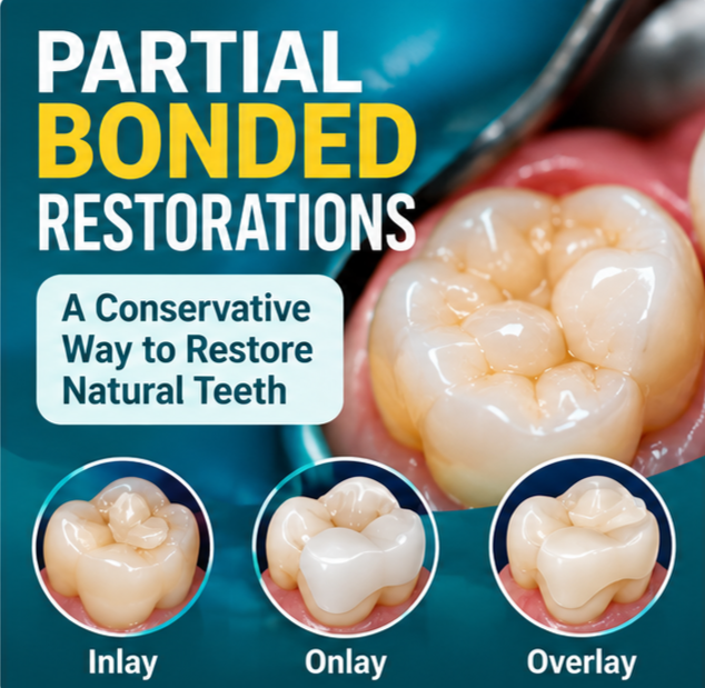
    Partial bonded restorations are conservative dental restorations used to repair damaged, weakened or extensively restored teeth while retaining as much healthy natural tooth structure as possible. Depending on the condition of the tooth, the restoration may replace only the damaged portion or cover one or more weakened cusps.

    Unlike a conventional full-coverage crown, which covers most or all of the visible clinical crown, a partial restoration is designed to cover only the areas that require reconstruction. It is bonded to the remaining tooth using an adhesive technique selected for the tooth and restorative material.

    Partial bonded restorations may be made from materials such as glass ceramics or resin composites. They are often considered as part of a conservative or biomimetic restorative approach, which aims to restore form and function while respecting the remaining natural tooth tissues.

    1. What Are Partial Bonded Restorations?

    Partial bonded restorations are custom-made or directly placed restorations that replace a portion of a tooth rather than covering the entire tooth. Common examples include inlays, onlays and overlays. The type selected depends on the location and extent of the damage, the condition of the remaining tooth structure and the patient’s bite.

    The main objectives are to:

    • Preserve healthy enamel and dentin wherever clinically possible.
    • Restore the tooth’s shape, contact points and chewing function.
    • Cover weakened cusps when additional protection is needed.
    • Provide a tooth-coloured result when an aesthetic material is appropriate.
    • Support a treatment plan tailored to the individual tooth.

    2. Why Is Preserving Natural Tooth Structure Important?

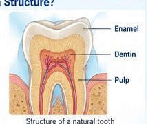
    A natural tooth contains several tissues, including enamel, dentin and the pulp. These structures work together during chewing. When decay, fracture, wear or an old restoration damages a tooth, treatment should address the problem while retaining sound tissue whenever possible.

    In suitable cases, a partial bonded restoration may avoid the need to prepare the entire circumference of the tooth for a full crown. However, conservative treatment is not appropriate in every situation. The dentist must assess the amount and quality of remaining tooth structure, the position of the defect, the margins available for bonding and the forces acting on the tooth.

    • Preservation of sound tooth structure: preparation can be limited to the areas that need treatment.
    • Adhesive retention: bonding can help connect the restoration to the remaining tooth when the conditions are suitable.
    • Restoration of function: the restoration can rebuild the chewing surface and tooth anatomy.
    • Aesthetic options: ceramic and composite materials can be selected to blend with natural teeth.

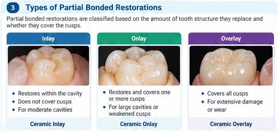
    3. Types of Partial Bonded Restorations

    A. Inlay Restoration

    An inlay fits within the prepared chewing surface of a tooth and does not cover its cusps. It may be considered when damage is contained within the central portion of the tooth and the cusps remain sufficiently strong.

    B. Onlay Restoration

    An onlay replaces a damaged portion of the tooth and covers one or more cusps when they need protection. It may be considered for teeth with larger cavities, weakened cusps or extensive existing restorations.

    C. Overlay Restoration

    An overlay covers the major cusps and a broader part of the chewing surface. It may be considered when there is extensive loss of occlusal tooth structure or significant wear, provided the remaining tooth and bonding conditions are suitable.

    The terms and designs can vary across clinical practices and materials. The dentist selects the restoration based on the tooth’s condition and the requirements of the chosen material.

    4. Biomimetic Dentistry and Partial Bonded Restorations

    Biomimetic dentistry is an approach that seeks to restore damaged teeth while preserving and making use of the remaining natural tissues. Partial bonded restorations can be part of this approach because they may combine conservative preparation with adhesive bonding.

    • Conservative preparation: remove decay and unsupported tissue while retaining sound structure where possible.
    • Appropriate adhesive technique: follow the bonding protocol for the tooth, restorative material and cement.
    • Cusp protection: cover weakened cusps when indicated by the clinical assessment.
    • Moisture control: isolate the working area appropriately to reduce contamination during bonding.
    • Functional assessment: check the restoration’s contacts and bite after placement.

    A bonded restoration does not reproduce every property of natural enamel and dentin. Its outcome depends on case selection, preparation design, material choice, adhesive procedures and maintenance.

    5. Who May Benefit from Partial Bonded Restorations?

    A dentist may consider a partial bonded restoration for a patient with:

    • A large cavity where a direct filling may not provide the required form or cusp protection.
    • A fractured or weakened cusp that needs coverage.
    • A defective or extensive existing restoration that requires replacement.
    • Tooth wear affecting the chewing surface.
    • A selected root canal-treated tooth where enough suitable tooth structure remains.
    • A need for a tooth-coloured restoration where the clinical conditions are favourable.

    Not every tooth is suitable. Extensive structural loss, deep or inaccessible margins, certain cracks, inadequate bonding conditions or complex bite problems may require another treatment. A clinical examination and any necessary diagnostic imaging are needed before deciding.

    6. Materials Used in Partial Bonded Restorations

    A. Glass-Ceramic Restorations

    Glass ceramics, including lithium disilicate, are used for selected indirect inlays, onlays and overlays. They can provide a natural-looking result and suitable mechanical properties when the design, thickness, bonding protocol and clinical indication are appropriate.

    B. Resin Composite Restorations

    Resin composite materials may be used for direct restorations or for selected indirect partial restorations. They are tooth-coloured and can be useful in cases where their material properties and repair options suit the clinical requirements.

    C. Other Restorative Materials

    Other ceramic and restorative materials may be considered according to the tooth, the restoration design and the available adhesive protocol. No single material is best for every patient. The dentist considers the amount of remaining enamel, tooth position, bite forces, aesthetic needs and the patient’s oral health.

    7. How Is the Procedure Performed?

    Step 1: Examination and treatment planning

    The dentist assesses the tooth, existing restoration, cracks, gum health, symptoms and bite. X-rays or other investigations may be recommended when needed.

    Step 2: Tooth preparation

    Decay and unsupported tooth structure are removed. The preparation is shaped according to the selected restoration and material, while preserving sound tissue where possible.

    Step 3: Impression or digital scan

    The prepared tooth is recorded using an intraoral scanner or a suitable conventional impression technique.

    Step 4: Restoration design and fabrication

    The restoration is designed to fit the tooth and reproduce its anatomy. It may be fabricated in a dental laboratory or through an appropriate digital workflow.

    Step 5: Try-in and fit assessment

    The dentist checks the fit, margins, contact points, appearance and relationship with opposing teeth. Adjustments are made when required.

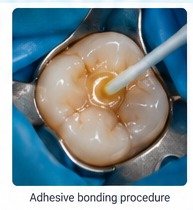
    Step 6: Adhesive bonding

    The tooth and restoration are treated according to the material-specific protocol. The restoration is then bonded using a suitable adhesive cementation technique, with appropriate isolation.

    Step 7: Bite adjustment and finishing

    The dentist checks the bite, makes any necessary adjustments and finishes or polishes the restoration.

    Step 8: Follow-up

    Review visits allow the dentist to assess the restoration, surrounding tissues and bite.

    8. Partial Bonded Restorations vs. Full Dental Crowns

    Both partial bonded restorations and full crowns can restore damaged teeth, but they differ in how much of the tooth they cover and how they are retained.

    FeaturePartial bonded restorationFull dental crown
    CoverageReplaces the damaged portion and may cover selected or all cusps.Covers most or all of the clinical crown.
    PreparationOften allows a more limited preparation when suitable.Usually requires preparation around the tooth.
    RetentionAdhesive bonding is central to retention.May rely on preparation geometry, cementation and, where suitable, adhesive bonding.
    Typical useWhen enough suitable tooth structure remains for a partial restoration.When broader coverage or a different retention strategy is required.

    Neither option is universally better. A partial bonded restoration may be suitable when sound tooth structure remains and reliable bonding is possible. A full crown may be more appropriate when the tooth needs broader coverage or the conditions do not favour a partial restoration.

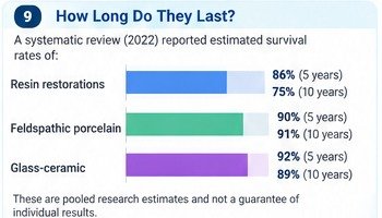
    9. How Long Do Partial Bonded Restorations Last?

    The lifespan of a partial bonded restoration varies. It is influenced by the restorative material, the amount of remaining tooth structure, the quality of bonding, bite forces, oral hygiene and regular maintenance.

    Published clinical studies report different survival outcomes for different materials and restoration types. Research estimates should not be treated as a guarantee for an individual patient. The dentist can discuss the expected performance and limitations of the proposed restoration based on the patient’s specific condition.

    Factors that may influence longevity include:

    • The amount and quality of remaining tooth structure.
    • Appropriate case selection and restoration design.
    • Moisture control and correct adhesive procedures.
    • Accurate fit, contacts and bite adjustment.
    • Good oral hygiene and regular dental reviews.
    • Assessment and management of clenching or grinding, when present.

    10. Potential Advantages

    • Conservative treatment: may preserve more healthy tooth structure than a full-coverage preparation in suitable cases.
    • Natural appearance: tooth-coloured materials can blend with surrounding teeth.
    • Customised reconstruction: the restoration can reproduce the tooth’s anatomy and contact relationships.
    • Cusp protection: selected weakened cusps can be covered when clinically indicated.
    • Adhesive retention: bonding can provide retention when the tooth and material are suitable.

    These benefits depend on clinical suitability and technique. Partial bonded restorations are not automatically the best choice for every damaged tooth.

    11. Aftercare: Maintaining Your Restoration

    • Brush twice daily with fluoride toothpaste.
    • Clean between the teeth daily using floss or an appropriate interdental cleaning aid.
    • Avoid using teeth to bite hard objects or open packaging.
    • If you grind or clench your teeth, ask your dentist whether a protective night guard is appropriate.
    • Attend recommended dental check-ups so the restoration and surrounding tissues can be monitored.
    • Contact your dentist if you develop persistent sensitivity, pain on biting, a change in your bite, or a loose or damaged restoration.

    12. Frequently Asked Questions

    Are partial bonded restorations better than dental crowns?

    They can be a conservative alternative when enough suitable tooth structure remains and reliable bonding is possible. Some teeth need full-coverage restorations. The right option depends on the clinical condition.

    Is the procedure painful?

    Local anaesthesia is used when required. Some patients may experience temporary sensitivity after treatment. Your dentist can explain what to expect in your case.

    Can a root canal-treated tooth receive a partial bonded restoration?

    Selected root canal-treated teeth can be restored with bonded onlays or overlays. The dentist must assess the remaining structure, cracks and functional loading before recommending this approach.

    Are partial bonded restorations made from ceramic?

    Ceramics such as glass ceramics are commonly used. Resin composite materials may also be suitable in selected cases.

    Can a partial bonded restoration break or come loose?

    Yes. Fracture, chipping, debonding and recurrent decay are possible complications. Case selection, bonding technique, bite assessment and maintenance are important.

    Are they suitable for worn teeth?

    They may be suitable for selected patients. The cause and severity of wear, available restorative space and bite must be assessed.

    Do they look natural?

    Tooth-coloured ceramic and composite materials can provide natural-looking results. Appearance depends on material, design, shade selection and clinical technique.

    How do I know whether I need an inlay, onlay, overlay or crown?

    A dental examination helps determine how much healthy tooth structure remains and whether any cusps need protection. The dentist can then recommend an appropriate option.

    13. Restorative Dental Care at Digital Bite Multispeciality Dental & Implant Hospital

    At Digital Bite Multispeciality Dental & Implant Hospital in Beeramguda, treatment planning is guided by the patient’s individual oral health needs. Where appropriate, modern diagnostic and digital dentistry technologies may support assessment and restorative planning.

    The clinical approach focuses on accurate diagnosis, appropriate material selection, restoration design and preservation of healthy tooth structure whenever possible. The most suitable treatment is determined after a dental examination.

    Book a Dental Consultation

    If you have a large filling, a fractured tooth, a worn chewing surface or an old restoration that needs assessment, arrange a dental consultation to find out which treatment may be suitable.

    DIGITAL BITE MULTISPECIALITY DENTAL & IMPLANT HOSPITAL

    ICRISAT Colony, Phase-1, Ameenpur Police Station Road, Beeramguda, Hyderabad
    Phone: 8019233440

    Preserve natural tooth structure. Restore function. Choose treatment tailored to your smile.

    14. References and Further Reading

    • Clinical guidelines for posterior restorations based on Coverage, Adhesion, Resistance, Esthetics, and Subgingival management (CARES concept): Part I – partial adhesive restorations. PubMed: https://pubmed.ncbi.nlm.nih.gov/37462378/
    • Comparative evaluation of clinical performance of ceramic and resin inlays, onlays, and overlays: A systematic review and meta-analysis. PubMed: https://pubmed.ncbi.nlm.nih.gov/36187858/
    • Survival of Partial Coverage Restorations on Posterior Teeth—A Scoping Review. PubMed Central: https://pmc.ncbi.nlm.nih.gov/articles/PMC12076103/

    Image and publishing checklist (for the website team)

    The grey image placeholders throughout this document indicate recommended image positions. Replace each placeholder with an original clinic photograph, a properly licensed stock image, or a clinically accurate illustration before publishing.

    • Use images that accurately represent the procedure and do not imply guaranteed results.
    • Obtain written patient consent before publishing identifiable clinical photographs.
    • Use descriptive alt text for accessibility and image SEO.
    • Compress images for faster page loading while retaining adequate quality.
    • Check the hospital name, address, phone number and all links before publishing.
    • Have a qualified dentist review the clinical content and references before publication.

    Suggested alt text examples:

    • Ceramic onlay restoration preserving natural tooth structure
    • Dental inlay, onlay and overlay restoration comparison
    • Adhesive bonding procedure for a partial dental restoration
    • Digital intraoral scan for a custom dental restoration

    Illustrative educational visuals. Individual treatment results and suitability vary.